Pancreatic Duct Blockage Risk Calculator
Your Risk Assessment
Risk Level:
Recommendations:
Important Note: This tool provides a general risk estimate. Consult with a healthcare provider for personalized medical advice.
Key Takeaways
- Pancreatic duct blockage prevents digestive enzymes from reaching the intestines, causing inflammation.
- Long‑standing blockage can create a cellular environment that favors tumor development.
- Common causes include gallstones, scar tissue (strictures), and benign tumors.
- Symptoms such as persistent abdominal pain, jaundice, and unexplained weight loss should prompt medical evaluation.
- Early imaging and blood‑marker testing dramatically improve chances of catching cancer before it spreads.
What Exactly Is Pancreatic Duct Blockage?
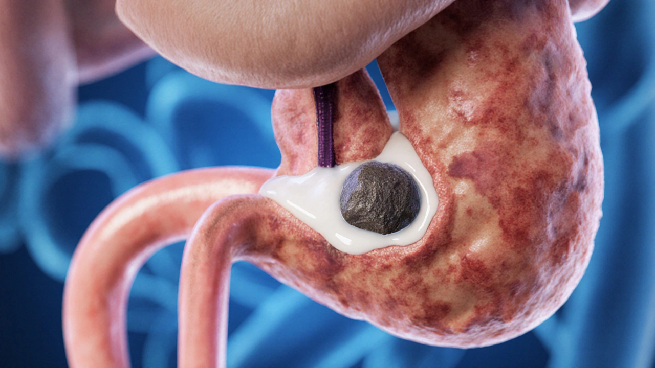
Pancreatic duct blockage is a condition where the main conduit that carries digestive enzymes from the pancreas to the small intestine becomes obstructed. The blockage can be partial or complete, and it often leads to a buildup of pancreatic fluid, which raises pressure inside the gland.
When the pressure climbs, the pancreas can become inflamed-a state known as pancreatitis. Over time, repeated inflammation creates scar tissue and alters the genetic makeup of the surrounding cells.
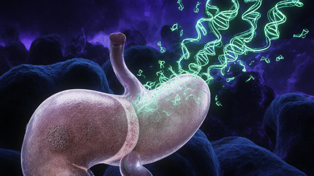
How Blockage Sets the Stage for pancreatic cancer
Scientists have identified several biological steps that link chronic duct obstruction to malignant transformation:
- Cellular stress: Stagnant fluid forces pancreatic cells to adapt to low‑oxygen conditions, triggering stress‑response pathways.
- DNA damage: Inflammatory enzymes release free radicals that can break DNA strands. Cells that survive may acquire mutations.
- Mutation accumulation: One of the most frequent mutations in pancreatic tumors affects the KRAS gene. Chronic inflammation raises the chance that KRAS‑mutated cells emerge.
- Proliferation signals: Damaged cells release growth factors that encourage nearby cells to divide, increasing the pool of potentially malignant cells.
- Immune evasion: Persistent inflammation can suppress normal immune surveillance, allowing abnormal cells to escape detection.
When enough of these steps line up, a benign lesion can evolve into an invasive tumor.
Common Causes of Duct Blockage
| Cause | Mechanism | Typical outcome if untreated |
|---|---|---|
| Gallstones | Stone migrates into the common bile duct and blocks the pancreatic duct entry point. | Acute pancreatitis, possible chronic blockage. |
| Strictures (scar tissue) | Repeated inflammation creates fibrotic rings that narrow the duct. | Persistent pain, progressive fibrosis, higher cancer risk. |
| Benign tumors (e.g., intraductal papillary mucinous neoplasm) | Growth inside the duct releases mucus that clogs the lumen. | Mucin buildup, chronic inflammation, malignant potential. |
| Pancreatic calcifications | Calcium deposits form from recurrent pancreatitis episodes. | Obstructive symptoms, increased tissue rigidity. |
Red‑Flag Symptoms That May Indicate a Blockage
Most people think of pancreatic problems as rare, but the warning signs are often subtle. Pay attention to:
- Steady, dull upper‑abdominal pain that worsens after meals. \n
- Yellowing of the skin or eyes (jaundice) when bile flow is compromised.
- Unexplained weight loss despite a normal appetite.
- Foul‑smelling, oily stools (steatorrhea) indicating enzyme deficiency.
- New‑onset diabetes or worsening blood‑sugar control.
If any of these persist for more than a few weeks, a medical work‑up is warranted.
How Doctors Diagnose a Blockage and Assess Cancer Risk
Imaging is the cornerstone of diagnosis. The most common tools include:
- CT scan - Provides cross‑sectional views and can spot stones, strictures, or masses.
- MRI/MRCP - Excellent for visualizing the ductal system without radiation.
- Endoscopic ultrasound (EUS) - Allows high‑resolution images and fine‑needle aspiration of suspicious tissue.
Blood tests complement imaging. The tumor marker CA 19‑9 is often elevated in pancreatic cancer, though it can rise with inflammation, too.
When imaging shows a blockage plus a suspicious lesion, doctors may recommend a biopsy via EUS‑guided needle to confirm malignancy.
Risk Factors That Amplify the Blockage‑to‑Cancer Pathway
Not everyone with a blocked duct develops cancer. Certain lifestyle and genetic factors stack the odds:
- Smoking: Increases KRAS mutation rates and reduces immune clearance.
- Heavy alcohol use: Drives chronic pancreatitis, a known precursor to cancer.
- Obesity: Alters hormone levels that can fuel tumor growth.
- Family history: Inherited mutations (e.g., BRCA2, PALB2) predispose the pancreas to malignant change.
- Diabetes: Both a risk factor and an early symptom of pancreatic disease.
Addressing these factors early can break the chain that leads from blockage to tumor.
Preventive Strategies and Early‑Detection Tips
While some risk elements (age, genetics) are out of our control, many actionable steps exist:
- Quit smoking - Even reducing pack‑years cuts mutation risk dramatically.
- Limit alcohol - Keep intake below the recommended 14 units per week.
- Maintain a healthy weight - Aim for a BMI under 25.
- Monitor abdominal pain - Keep a diary; seek care if pain is persistent or radiates to the back.
- Regular medical check‑ups - High‑risk individuals (family history, chronic pancreatitis) should discuss screening MRI or EUS with their gastroenterologist.
Early detection dramatically improves survival; localized pancreatic cancer has a 5‑year survival rate above 30%, versus less than 5% once it spreads.
Treatment Options When Blockage Has Already Triggered Cancer
Management depends on stage:
- Surgery - The Whipple procedure (pancreaticoduodenectomy) removes the head of the pancreas, surrounding tissue, and the obstructed duct. It offers the best chance for cure when the tumor is confined.
- Chemotherapy - Regimens such as FOLFIRINOX or gemcitabine plus nab‑paclitaxel shrink tumors and target microscopic disease.
- Radiation therapy - Often combined with chemo to improve local control.
- Palliative stenting - Endoscopic placement of a metal stent can relieve duct obstruction, reducing pain and jaundice.
Multidisciplinary care-surgeons, oncologists, gastroenterologists, dietitians-yields the best outcomes.
Living With the Aftermath: Lifestyle Adjustments Post‑Treatment
Recovery focuses on nutrition, enzyme replacement, and monitoring for recurrence.
- Pancreatic enzyme supplements aid digestion and prevent malabsorption.
- Low‑fat diet reduces stress on the remaining pancreatic tissue.
- Regular follow‑up imaging (usually every 3‑6 months) catches any regrowth early.
- Physical activity supports metabolic health and can improve blood‑sugar control for those who develop diabetes.
Emotional support-counseling groups or peer networks-helps patients cope with the psychological impact of a cancer diagnosis.
Frequently Asked Questions
Can a single episode of pancreatitis cause pancreatic cancer?
A one‑time bout of acute pancreatitis usually resolves without long‑term risk. It's the repeated or chronic inflammation that raises the chance of cancer.
Is there a screening test for people with pancreatic duct blockage?
Screening isn’t routine for the general population, but high‑risk individuals may undergo MRI/MRCP or endoscopic ultrasound every 1‑2 years.
How effective are stents at relieving blockage pain?
Metal stents often relieve pain and jaundice within days, allowing patients to resume normal diet while other treatments are planned.
What lifestyle changes lower my risk after a blockage is treated?
Quit smoking, limit alcohol, maintain a healthy weight, and follow a low‑fat diet with enzyme supplements as prescribed.
Does a high CA 19‑9 level always mean cancer?
No. CA 19‑9 can rise with inflammation, gallstones, or liver disease. It’s useful when combined with imaging and clinical context.


Jeremy Olson
October 4, 2025 AT 14:32Pancreatic duct obstruction is more than a mechanical issue; it creates a chronic inflammatory environment that can promote malignant transformation. Studies show that prolonged blockage increases the risk of acinar cell injury, fibrosis, and dysplasia. Early detection through imaging and functional tests can guide timely intervention, potentially lowering cancer incidence.
Ada Lusardi
October 5, 2025 AT 00:16I'm scared, but grateful for the calculator! 😊
Pam Mickelson
October 5, 2025 AT 09:42The risk algorithm you posted accounts for many known contributors, such as age and smoking status. However, remember that BMI and alcohol intake interact synergistically, amplifying the inflammatory cascade. Adding a family-history weighting for first-degree relatives could improve predictive accuracy.
Joe V
October 5, 2025 AT 19:42Oh great, another checklist that pretends to solve a problem that needs a surgeon’s scalpel. The calculator is useful for self‑assessment, but it won’t replace a proper endoscopic evaluation. Still, kudos for making the risk factors visible to the layperson.
Scott Davis
October 6, 2025 AT 04:36Short and sweet: blockages over six months boost risk. Get checked.
Calvin Smith
October 6, 2025 AT 15:09Listen up, folks-if you’ve got a blocked duct and you’re also lighting up cigarettes, you’re practically handing cancer the keys to your pancreas. Add a splash of heavy drinking, and you’ve got a recipe for disaster. The calculator’s point system is a neat gimmick, but the reality is blunt: eliminate those risk habits or face the consequences. Doctors love numbers, but they love healthy patients even more.
Brenda Hampton
October 6, 2025 AT 23:29The interactive tool is a great way to engage people who might otherwise ignore asymptomatic risk factors. By visualizing the score, users can see how lifestyle tweaks lower the total. It also encourages conversations with primary‑care physicians, which is essential for early imaging. Keep iterating on the UI; a smoother flow will boost compliance.
Lara A.
October 7, 2025 AT 08:56Wow-look at this, another “medical calculator” that pretends to predict cancer!;;; The pharma industry loves these gimmicks; they push fear to sell drugs. Remember, the real danger is hidden in the data they keep from us.
Ashishkumar Jain
October 7, 2025 AT 18:39Jeremy, you nailed the biology-chronic inflammation is the silent driver. In my experience, patients who get a CT scan after a six‑month blockage often have precancerous lesions that are catchable. Also, incorporating serum CA19‑9 levels could add another layer of detection. Keep spreading the word; awareness saves lives.
Gayatri Potdar
October 8, 2025 AT 04:39Ada, the calculator looks innocent but it’s part of a larger agenda to keep us scared. They want us to buy expensive monitoring kits while ignoring natural prevention. Don’t be fooled-focus on diet and movement, not on a web form.
Marcella Kennedy
October 8, 2025 AT 13:49Pam, your concise summary is appreciated, and I’d love to expand on a few points that merit deeper discussion. First, the role of chronic pancreatitis as a precursor to adenocarcinoma is well‑documented; repeated episodes of inflammation lead to cellular turnover, DNA damage, and eventually malignant transformation. Second, emerging data suggest that certain genetic polymorphisms, such as BRCA2 and PALB2 mutations, significantly elevate risk when combined with environmental factors like smoking and alcohol abuse. Third, the gut microbiome’s influence on pancreatic inflammation is an exciting frontier-dysbiosis may exacerbate fibrosis and create a pro‑tumorigenic microenvironment, making microbiome modulation a potential preventive strategy.
Moreover, the risk calculator you shared could benefit from incorporating a weighted score for familial cancer syndromes, as hereditary pancreatitis carries a markedly higher lifetime incidence. Including a question about known pathogenic mutations would personalize risk stratification further. Fourth, imaging modalities matter: endoscopic ultrasound (EUS) has higher sensitivity for detecting early ductal changes compared to conventional CT, especially in patients with borderline scores. Early referral for EUS in high‑risk individuals can catch precancerous lesions before they become surgically unresectable.
Finally, lifestyle interventions remain the cornerstone of risk reduction. Smoking cessation reduces the relative risk by up to 40% within five years, while maintaining a BMI under 25 and limiting alcohol intake to under 14 units per week further diminish the inflammatory burden. Patients who adopt these changes also tend to have better overall metabolic health, which indirectly protects pancreatic tissue from oxidative stress.
In summary, pancreatic duct blockage is a multifactorial problem; integrating genetics, microbiome insights, advanced imaging, and aggressive lifestyle modification into the calculator will enhance its predictive power and, most importantly, translate into real‑world reductions in pancreatic cancer incidence.
Jamie Hogan
October 9, 2025 AT 00:06One might argue the algorithm is a mere approximation, yet it does provide a structured framework for risk appraisal.
Ram Dwivedi
October 9, 2025 AT 09:32Great points, Ashish! Adding a note about the utility of MRCP for non‑invasive duct imaging can help patients avoid unnecessary ERCPs. Also, the inclusion of pancreatic enzyme supplementation for chronic obstruction may improve quality of life while we monitor for malignancy. 😊
pooja shukla
October 9, 2025 AT 19:16Our nation’s health policies ignore the real culprits-Western diets and sedentary habits. The calculator is a wake‑up call that our people need to take charge and reject imported junk. If we don’t act, we’ll be paying for foreign treatment and pharmaceutical profits.
Poonam Mali
October 10, 2025 AT 04:09Poora, the pathophysiology is clear: cytokine storms, desmoplastic reaction, and oncogenic signaling converge. Your patriotism won’t cure the molecular cascade.
Alan Whittaker
October 10, 2025 AT 14:09They don’t want you to know that most studies are funded by the same conglomerates that profit from the very diseases they claim to fight. Keep your eyes open.
Michael Waddington
October 10, 2025 AT 23:36Sounds like a typical scare‑tactic to sell supplements. Not convinced.
HAMZA JAAN
October 11, 2025 AT 08:46Scott, your brief warning hits the mark-duration matters. A follow‑up scan after three months is prudent.
April Rios
October 11, 2025 AT 18:29The calculator’s scoring system reflects epidemiological data, yet it fails to capture the nuanced interplay of epigenetics. A truly comprehensive model would integrate methylation markers alongside lifestyle inputs. That said, it’s a solid foundation for public awareness.
Natasha Beynon
October 12, 2025 AT 03:56April, I appreciate your balanced critique. Incorporating emerging biomarkers, as you suggested, could indeed refine risk stratification. Meanwhile, encouraging patients to discuss the tool with their gastroenterologists ensures it’s used responsibly.